Welcome to the Organoid Zoo!
In our lab, we grow 3D models of the placenta called organoids from a wide range of mammals, including humans, monkeys, and even bats. These living models let us explore the inner workings of pregnancy in ways that were never before possible. Each organoid in our “zoo” represents a different species and reveals how evolution has shaped the placenta to meet unique reproductive challenges. Some placentas must function during flight, others in long gestations, or in environments full of pathogens. Organoids help us uncover how these systems work, and how they sometimes fail. By studying these differences side by side, we’re gaining new insight into how the placenta grows, protects, and supports life across species—including our own.
Learn about some of our models below!
Meet the Rhesus Macaque: One of Our Closest Cousins
Rhesus macaques share over 90% of their DNA with humans, making them one of our closest evolutionary relatives. But when it comes to pregnancy, even small genetic differences can lead to big biological changes. In fact, the structure of the rhesus macaque placenta is distinct from that of humans, with differences in how cells organize, interact, and support fetal development. The specialized cells that build the macaque placenta are uniquely adapted to their species and we’re just beginning to understand how. To investigate these differences, we’ve created rhesus macaque placental organoids. These living systems allow us to study how the placenta grows, communicates with the fetus, and defends against infections in a species-specific way. By comparing organoids from macaques, humans, and other mammals, we’re uncovering both shared foundations and evolutionary innovations in how life begins. Read about our rhesus macaque models here.
Organoids and Spatial Maps: Exploring the Pig Placenta Cell by Cell
Pigs may not be our closest relatives, but their pregnancies offer a fascinating contrast to our own. Unlike humans, pigs have a structurally distinct placenta—one that supports multiple fetuses simultaneously, with separate zones for nutrient exchange and immune regulation. Despite these differences, pigs still share key features with human pregnancy, making them a powerful model for studying fetal development, maternal health, and infection. What makes pigs especially valuable is our ability to study their placentas with extraordinary precision. Using advanced tools like placental organoids and spatial transcriptomics, we can now map gene activity across the pig placenta, cell by cell, and region by region, to understand how it grows, adapts, and defends developing fetuses.
These tools let us ask big questions:
– How does the placenta balance nutrients and immune protection for an entire litter?
– What happens when these systems break down?
– And how can we apply these insights to better understand and protect human pregnancies?
By combining cutting-edge technology with comparative biology, our work in pigs helps reveal the core principles of pregnancy—and the remarkable ways evolution has solved the same challenge in different species. Read more about our pig models here.
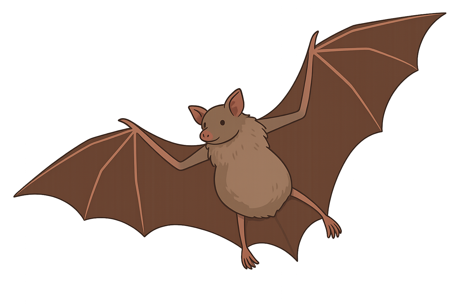
What Makes a Bat Placenta So Strange—and So Smart?
Bats may seem worlds apart from humans, but they offer extraordinary insights into how pregnancy can work under extreme conditions. Despite being one of the most evolutionarily distant mammals we study, bats have evolved complex placentas that support long gestations, high fetal investment, and constant exposure to pathogens—all while flying. What makes bat pregnancy even more fascinating is that their placental structure and cell types are strikingly different from those in humans and primates. These unique features may help explain why bats can tolerate viral infections that cause harm in other species, including humans. To explore these adaptations, we grow bat placental organoids. These organoids allow us to study how bat placentas develop, function, and protect the fetus. By comparing them to human, mouse, and primate models, we’re uncovering how evolution has shaped distinct, but equally effective, solutions to supporting life before birth. Read more about our bat models here.


